Biology of the schizont: Difference between revisions
From haematologyetc.co.uk
No edit summary |
No edit summary |
||
| (19 intermediate revisions by the same user not shown) | |||
| Line 3: | Line 3: | ||
<span style="font-size:80%">(click blue highlighted text to return to page)</span></br></br> | <span style="font-size:80%">(click blue highlighted text to return to page)</span></br></br> | ||
<span style="font-size:90%">[[Malaria Index|Malaria main index]]</span></br> | <span style="font-size:90%">[[Malaria Index|Malaria main index]]</span></br> | ||
<span style="font-size:90%">>[[Malaria Biology| | <span style="font-size:90%">>[[Malaria Biology|Malaria biology]]</span></br> | ||
---- | ---- | ||
| Line 27: | Line 15: | ||
<gallery mode="nolines" widths=300px heights=600px> | <gallery mode="nolines" widths=300px heights=600px> | ||
File:Blood_stages_schizonts_only.jpg|<span style="font-size:80%">''Basic | File:Blood_stages_schizonts_only.jpg|<span style="font-size:80%">''Basic schizont development (coloured images)''</span>|link={{filepath:Blood_stages_schizonts_only.jpg}} | ||
</gallery> | </gallery> | ||
| Line 44: | Line 32: | ||
(2) This is followed by further cycles of replication </br> | (2) This is followed by further cycles of replication </br> | ||
(3) In this case this results in the formation of 8 daughter parasites </br> | (3) In this case this results in the formation of 8 daughter parasites </br> | ||
(4) The daughter parasites mature and the red cell ruptures to | (4) The daughter parasites mature and the red cell ruptures to release the “merozoites” </br> | ||
(5) The released merozoites very rapidly infect new red cells (so rapid that free merozoites will not usually be seen in blood). | (5) The released merozoites very rapidly infect new red cells (so rapid that free merozoites will not usually be seen in blood). | ||
| Line 50: | Line 38: | ||
{| class="wikitable" style="border-style: solid; border-width: 5px; border-color: #023020; color:black" | {| class="wikitable" style="border-style: solid; border-width: 5px; border-color: #023020; color:black" | ||
|colspan="1" style = "font-size:100%; color:black; background: #afbddb |'''Morphological features | |colspan="1" style = "font-size:100%; color:black; background: #afbddb |'''Morphological features and relevance''' | ||
|} | |} | ||
(1) '''The number of replication cycles differs between species:''' the typical number of merozoites formed differs between species with as few as 8 (in P.malariae) up to a possible 32 (in P.vivax)</br> | (1) '''The number of replication cycles differs between species:''' the typical number of merozoites formed differs between species with as few as 8 (in P.malariae) up to a possible 32 (in P.vivax)</br> | ||
(2) '''This stage may not always | (2) '''This stage may not always occur in blood:''' schizonts of ''P.falciparum'' adhere within the small vessels so is not seen in blood unless infection is very severe | ||
</br> | </br></br> | ||
<gallery mode="nolines" widths="200px" heights="220px" > | |||
File:Schizontreal4.jpg|Mature schizont releasing merozoites|link={{filepath:Schizontreal4.jpg}} | |||
</gallery> | |||
<span style="font-size:200%">→</span> [[Click for images of schizont morphology|Click for clinical images illustrating schizont development]] | |||
-------- | ---- | ||
Relevance of schizonts to | |||
{| class="wikitable" style="border-style: solid; border-width: 5px; border-color: #023020; color:black" | |||
|colspan="1" style = "font-size:100%; color:black; background: #afbddb |'''Relevance of schizonts to clinical biology''' | |||
|} | |||
The release of merozoites from schizonts exposes the body to large amounts of free parasite antigens no longer contained within the erythrocytes - the result is an immune response causing high fever and illness symptoms. In some cases the development of parasites is synchronous so that all schizonts mature and release their merozoites at the same time - although rarely seen now, this pattern of development may produce a pattern of remitting fever with a distinct periodicity depending on species: underlying the older descriptive terms tertian or quartan malaria. | |||
---- | |||
Latest revision as of 14:02, 31 May 2024
Navigation
(click blue highlighted text to return to page)
Malaria main index
>Malaria biology
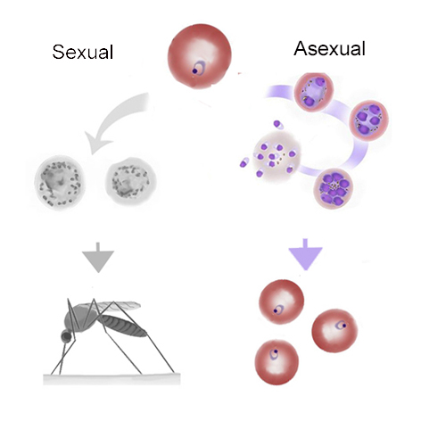
| The schizont pathway |
The stage begins with the first cycle of asexual replication forming a recognisable “schizont” then concludes when the individual “merozoites” are released to infect new erythrocytes.

Basic schizont development (coloured images)
| Merozoite release |

Formation and release of merozoites
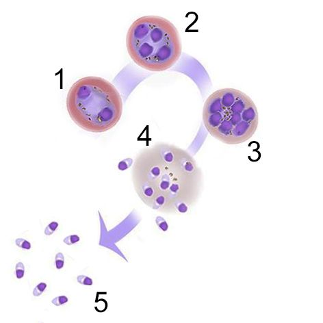
(1) The stage begins with the first cycle of asexual division producing two chromatin masses
(2) This is followed by further cycles of replication
(3) In this case this results in the formation of 8 daughter parasites
(4) The daughter parasites mature and the red cell ruptures to release the “merozoites”
(5) The released merozoites very rapidly infect new red cells (so rapid that free merozoites will not usually be seen in blood).
| Morphological features and relevance |
(1) The number of replication cycles differs between species: the typical number of merozoites formed differs between species with as few as 8 (in P.malariae) up to a possible 32 (in P.vivax)
(2) This stage may not always occur in blood: schizonts of P.falciparum adhere within the small vessels so is not seen in blood unless infection is very severe
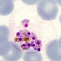
Mature schizont releasing merozoites

→ Click for clinical images illustrating schizont development
| Relevance of schizonts to clinical biology |
The release of merozoites from schizonts exposes the body to large amounts of free parasite antigens no longer contained within the erythrocytes - the result is an immune response causing high fever and illness symptoms. In some cases the development of parasites is synchronous so that all schizonts mature and release their merozoites at the same time - although rarely seen now, this pattern of development may produce a pattern of remitting fever with a distinct periodicity depending on species: underlying the older descriptive terms tertian or quartan malaria.