Gallery of early trophozoites: Difference between revisions
From haematologyetc.co.uk
No edit summary |
No edit summary |
||
| Line 45: | Line 45: | ||
---- | ---- | ||
<span style="font-size:95%">''' ''P.knowlesi'' '''</span></br> | <span style="font-size:95%">''' ''P.knowlesi'' '''</span></br> | ||
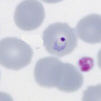
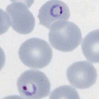
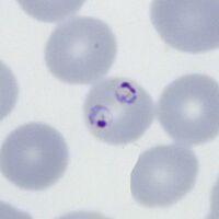
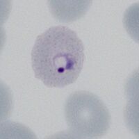
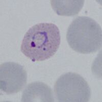
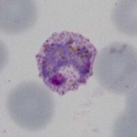
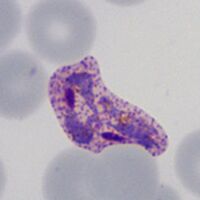
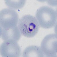
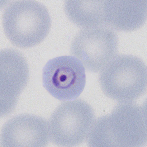
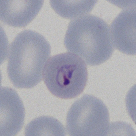
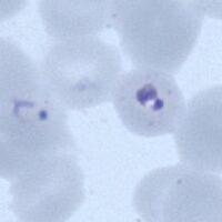
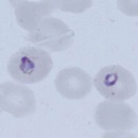
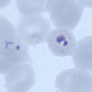
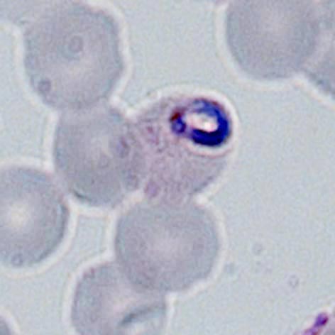
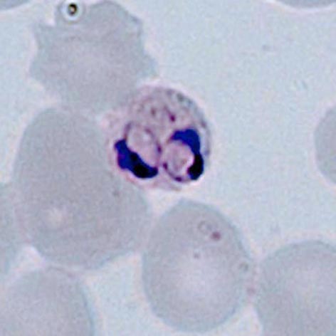
<span style="font-size:95%">At the early trophozoite stage an infection by ''P.knowlesi'' resembles that by ''P.falciparum'' and may include cells that have double chromatin dots, muliply infected red cells, and | <span style="font-size:95%">At the early trophozoite stage an infection by ''P.knowlesi'' resembles that by ''P.falciparum'' and may include cells that have double chromatin dots, muliply infected red cells, and accolé forms. This may create diagnostic confusion in cases where only early trophozoites are present. Later forms begin to resemple parasites of ''P.malariae'' and hese should be specifically sought where infections arise in geographical areas associated with this parasite. | ||
<gallery mode="traditional" widths=200px heights=200px> | <gallery mode="traditional" widths=200px heights=200px> | ||
File:PKET1.jpg|<span style="font-size:80%"> | File:PKET1.jpg|<span style="font-size:80%">Fine early rings</span>|link={{filepath:PKET1.jpg}} | ||
File:PKET2a.jpg|<span style="font-size:80%"> | File:PKET2a.jpg|<span style="font-size:80%">Double dot/fimbriation</span>|link={{filepath:PKET2a.jpg}} | ||
File:PKET3a.jpg|<span style="font-size:80%"> | File:PKET3a.jpg|<span style="font-size:80%">Accolé form/fimbriation</span>|link={{filepath:PKET3a.jpg}} | ||
File:PKET4a.jpg|<span style="font-size:80%"> | File:PKET4a.jpg|<span style="font-size:80%">Multiple infection</span>|link={{filepath:PKET4a.jpg}} | ||
</gallery>" | </gallery>" | ||
---- | ---- | ||
Revision as of 11:06, 3 June 2024
Navigation
Go Back
P.falciparum
Small delicate rings, within red cells of normal (or slightly crenated) appearance.
These may be the only forms seen in some patients at diagnosis.
Some parasite forms are typical though not exclusive of the species, these include: accolé forms, double chromatin dot forms, and multiple parasites within infected red cells.
Fine ring form
Double dot form and normal ring

Accolé and double dot forms
Multiple parasite form


"
P.vivax
Rings begin as small forms in normal sized red cells, but as they develop both parasites and red cells become markedly enlarged and irregular. Schuffners dots develop during this stage initially as a fine dusting but becoming more prominent.
Early ring form
Early ring form
Intermediate trophozoite
Intermediate/late trophozoite


"
P.ovale
As with the other species development begins with small forms and normal red cells; However as the species develops cahnges to red cells begin that might include finbriation, ovoid form and some enlargmnet. Similar to P.vivax the cytoplasmic James (Schuffners) dots appear initially as a fine dots but becoming more prominent as the parasites mature.
Early ring form

Ring with dots/fimbriation
faint Ziemann's dots

Ring early ovoid change

"
P.malariae
Generally parasites are infrequent. The very early small forms become a little more robust than P.falciparum, and may acquire features more typical (though not exclusive) for species including central chromatin dot forms, and early parasite elongation or angular forms. Red cells have normal size and shape or may have reduced size, cytoplasmic dots should not be present (although the uncommon fine Stinton's dots may be seen).
Ring form in small red cell
The central chromatin dot

Early elongation, Stinton's dots
Early angularity of form
"
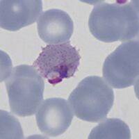
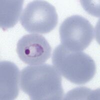
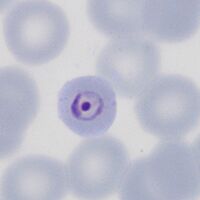
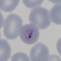
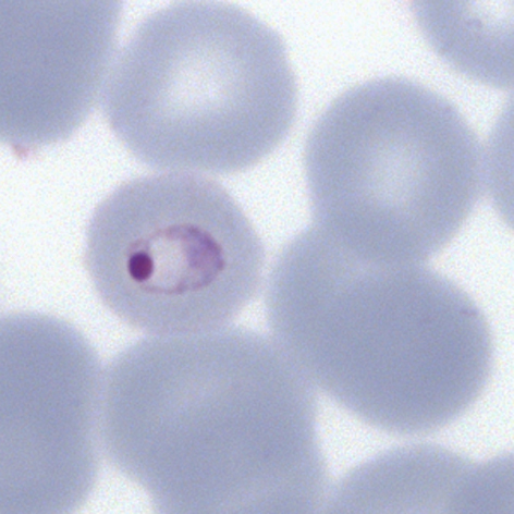
P.knowlesi
At the early trophozoite stage an infection by P.knowlesi resembles that by P.falciparum and may include cells that have double chromatin dots, muliply infected red cells, and accolé forms. This may create diagnostic confusion in cases where only early trophozoites are present. Later forms begin to resemple parasites of P.malariae and hese should be specifically sought where infections arise in geographical areas associated with this parasite.
Fine early rings
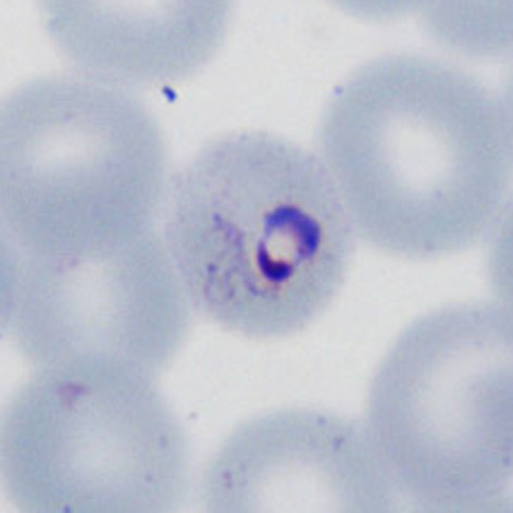
Double dot/fimbriation

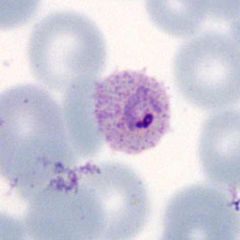
Accolé form/fimbriation

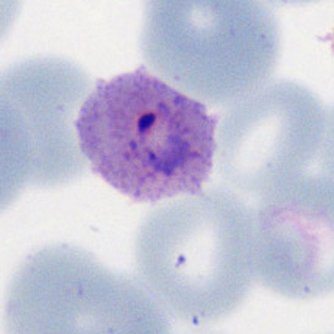
Multiple infection

"