Gallery of early trophozoites: Difference between revisions
From haematologyetc.co.uk
No edit summary |
No edit summary |
||
| Line 18: | Line 18: | ||
<gallery mode="traditional" widths=200px heights=200px> | <gallery mode="traditional" widths=200px heights=200px> | ||
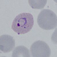
File:PVET1g.jpg|<span style="font-size:80%"> | File:PVET1g.jpg|<span style="font-size:80%">Very early double dot</span>|link={{filepath:PVET1g.jpg}} | ||
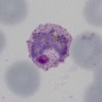
File:PVET2g.jpg|<span style="font-size:80%">Double dot form and normal ring</span>|link={{filepath:PVET2g.jpg}} | File:PVET2g.jpg|<span style="font-size:80%">Double dot form and normal ring</span>|link={{filepath:PVET2g.jpg}} | ||
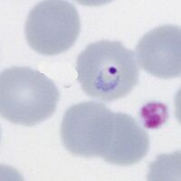
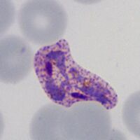
File:PVET3g.jpg|<span style="font-size:80%">Accolé and double dot forms</span>|link={{filepath:PVET3g.jpg}} | File:PVET3g.jpg|<span style="font-size:80%">Accolé and double dot forms</span>|link={{filepath:PVET3g.jpg}} | ||
Revision as of 22:27, 22 May 2024
Navigation
Go Back
P.falciparum
Small delicate rings, within red cells of normal (or slightly crenated) appearance. Some parasite forms are typical though not exclusive of the species, these include: accolé forms, double chromatin dot forms, and multiple parasites within infected red cells.
Fine ring form
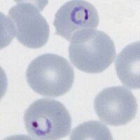
Double dot form and normal ring

Accolé and double dot forms
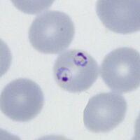
Multiple parasite form


"
P.vivax
Rings begin as small forms in normal sized red cells, but as they develop both parasites and red cells become larger and irregular. Schuffners dots develop during this stage initially as a fine dusting but becoming more prominent.
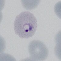
Very early double dot
Double dot form and normal ring
Accolé and double dot forms
Multiple parasite form


"