Gallery of early trophozoites: Difference between revisions
From haematologyetc.co.uk
No edit summary |
No edit summary |
||
| Line 15: | Line 15: | ||
---- | ---- | ||
<gallery mode="traditional" widths=240px heights= | <gallery mode="traditional" widths=240px heights=180px> | ||
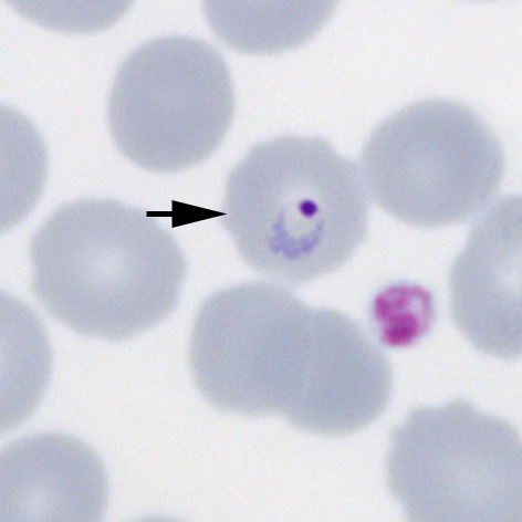
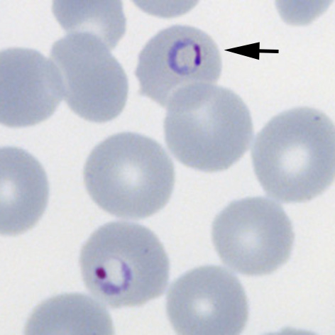
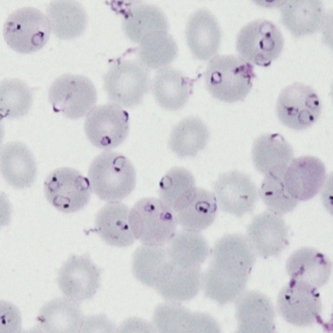
File:PFET1p.jpg|<span style="font-size:80%">'''Fine ring form''' The small and delicate form of this species</span>|link={{filepath:PFET1p.jpg}} | File:PFET1p.jpg|<span style="font-size:80%">'''Fine ring form''' The small and delicate form of this species</span>|link={{filepath:PFET1p.jpg}} | ||
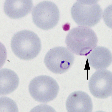
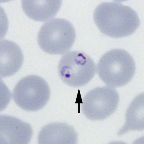
File:PFET2p.jpg|<span style="font-size:80%">'''Double chromatin dot form''' Two chromatin dots (sometimes known as "signet ring" form).</span>|link={{filepath:PFET2p.jpg}} | File:PFET2p.jpg|<span style="font-size:80%">'''Double chromatin dot form''' Two chromatin dots (sometimes known as "signet ring" form).</span>|link={{filepath:PFET2p.jpg}} | ||
Revision as of 21:25, 22 May 2024
Navigation
Go Back
|