Morphology of the sexual phase of parasite development
From haematologyetc.co.uk
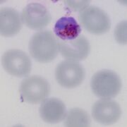
A
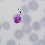
B
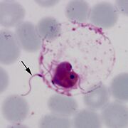
C
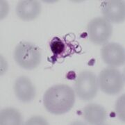
D
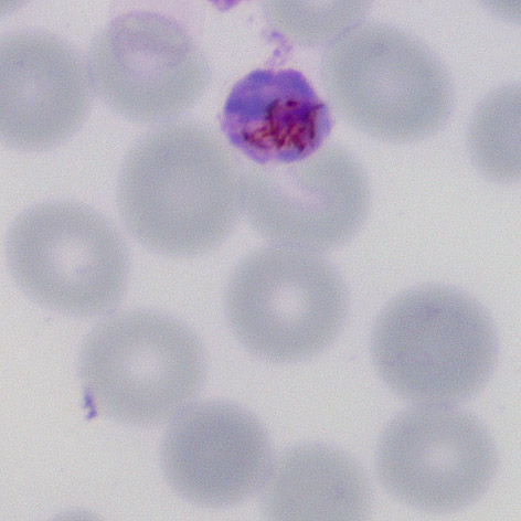

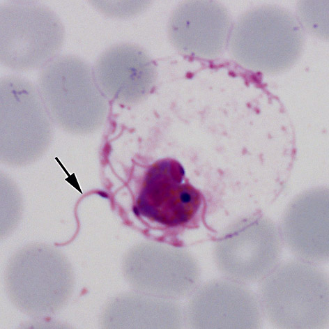
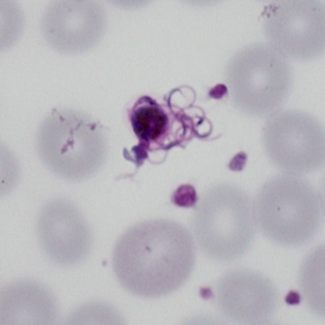
In the initial stage involves both the male microgametocytes and female macrogametocytes swelling and becoming more globular. The stage shown in (A) may represent an oocyte although it is not certain (these often clump together). The following stages are quite recognisable for the male gametocte as the male gametes burst from the erythrocytes in the process of exflagellation this may be seen as the early emergence (B), red cell swelling and disollution (C) and emergence of the gametes (D).